A series of inherited eye conditions known as juvenile macular dystrophy (also known as juvenile macular degeneration) affects children and young adults. Age-related macular degeneration (ADM) and juvenile macular dystrophy are distinct conditions. Although AMD is a natural aspect of ageing, juvenile macular degeneration is a hereditary disease (passed down from parents).
A small region of the retina is called the macula. Even though it only makes up a small portion of the retina, it is significantly more detail-sensitive than the rest of the retina. Your macula provides you with your central vision and enables you to clearly perceive little details. You can thread a needle, read small print, and read road signs because to the macula.
Stargardt disease is the most prevalent type of juvenile macular dystrophy. Juvenile retinoschisis and Best’s disease, often known as Best’s vitelliform retinal dystrophy, are other forms of juvenile macular dystrophy.
These uncommon diseases all result in loss of central vision.
Juvenile macular dystrophy comes in many different forms, yet they are all comparable. All of these interfere with your central vision. Your central vision could be blurry, distorted, or have black spots. In most cases, side vision is unaffected, although in the later stages, colour perception may impaired. Children or teenagers may experience the initial symptoms. Each eye is not usually equally affected by these symptoms.
Juvenile macular dystrophy can cause some people to retain functional vision well into adulthood. Others experience a faster rate of disease progression. Best’s disease patients frequently have eyesight that is virtually normal for many years. It is possible that many people are not even aware they have it. In contrast, Stargardt disease frequently causes 20/200 vision. which is characterized as legal blindness.
A hereditary condition that runs in the family is juvenile macular dystrophy. Accordingly, the disease is inherited from parents to children. Different disease kinds have various modes of inheritance.
For instance, Stargardt disease typically has a recessive gene. This implies that for the disease to manifest, it must be inherited from both parents. The disease is most frequently “carried” by parents. This means they do not have the disease but can silently pass the diseases to their children.
A dominant gene causes Best’s disease. This indicates that a child can become ill by inheriting the gene from just one parent. There is a 50% (5 out of 10) probability that a child born to an affected person and an unaffected partner will contract the disease.
Males are disproportionately affected by the X-linked condition known as juvenile retinoschisis. The X chromosome is home to the disease-causing genetic mutation. This chromosome is passed down to males from their mothers. The Y chromosome comes from fathers.
To examine the retina, your ophthalmologist will do a dilated eye exam. Juvenile macular dystrophy patients exhibit symptoms unique to their condition.
Yellowish specks in and under the macula are possible in those who have Stargardt disease. The specks are deposits of lipofuscin, an abnormally accumulating fatty material in people with Stargardt disease.
Under the macula, a yellow cyst develops in Best’s disease sufferers. When the cyst eventually bursts, fluid and yellow deposits are released. These might cause macula damage.
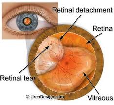
The macula is impacted by the retina splitting into two layers in juvenile retinoschisis. Blisters may develop in the gaps between these layers. The vitreous, the liquid that fills the eye, is susceptible to blood vessel leakage. Retinal detachments may result from juvenile retinoschisis.
To confirm the diagnosis, your ophthalmologist could do an OCT or fluorescein angiography, a laser scan of the retina. A dye is injected into your arm during this test. As the dye moves through the blood vessels in your retina, a picture of it is taken. ERG (electroretinography) testing may also be recommended by your ophthalmologist. The retina’s electrical activity is assessed during this test. Genetic testing is currently done in specialised labs. They are able to identify the precise gene responsible for the illness. Knowing this is crucial since different genes can result in the same retinal appearance.
Juvenile macular degeneration is still incurable. But numerous clinical trials for gene therapy are currently being conducted. It is possible to treat the faulty gene in the retina with gene therapy. These therapies might be able to halt the disease’s progression and stop visual loss.
Sunglasses can help shield the eyes from UV rays and strong light. Mobility training and low vision equipment can help patients cope with their visual loss. The importance of genetic testing through your ophthalmologist cannot be overstated. This identifies the specific gene responsible for your macular dystrophy.
At The Eye Center- Dr. Mahnaz Naveed Shah & Associates our team of eight ophthalmology subspecialists/ eye specialists, eye surgeons who are considered amongst the very best eye specialists in Karachi and in Pakistan, have the diagnostic and treatment capabilities to treat from the simplest to the most complex patients. We work hard to provide our patients with the best possible medical and surgical eye care, in a state of the art purpose built eye care facility. We offer the entire array of medical, laser and surgical treatments to help provide patients the best possible care in the most efficient, safe and ethical manner.
If you need an appointment, please contact us at 03041119544 during our working hours or leave us a WhatsApp message at +923028291799 and someone will connect with you. Walk-in appointments are also available for emergencies. We can also be reached through our web portal on www.surgicaleyecenter.org

