Our Services
Our Services
A Full Spectrum of Vision Services, Tailored to Every Patient's Needs
At The Eye Center – Dr. Mahnaz Naveed Shah & Associates, we offer a complete range of eye care services tailored to meet the needs of every patient. From routine checkups to advanced diagnostics and surgical treatments, our experienced team is here to protect, preserve, and enhance your vision — with compassion, precision, and care you can trust.
Explore our key services below: possible.

At The Eye Center we provide clinical and surgical expertise in all areas of subspecialty eye care. Our nine general and sub-specialist ophthalmologists have been extensively trained and experienced in providing a diverse array of routine and complex treatment to adults as well as to children.
Our eye specialists & eye surgeons are counted amongst the best within the city and are considered well recognised experts within their respective areas of subspecialty interests all over Pakistan.
Our 14 eye specialist share the same medical record for the patients who have been seen at The Eye Center and have access to all digital imaging that has been carried out for a particular patient. This makes it far more convenient for a patients who are seeing us for complex clinical care to have a continuous follow-up and have that follow-up documented within the medical records.
Should any of the doctors be unavailable at the time of an emergency, others doctors within our team communicate with the primary ophthalmologist for that patient hence ensuring uninterrupted and optimal care.
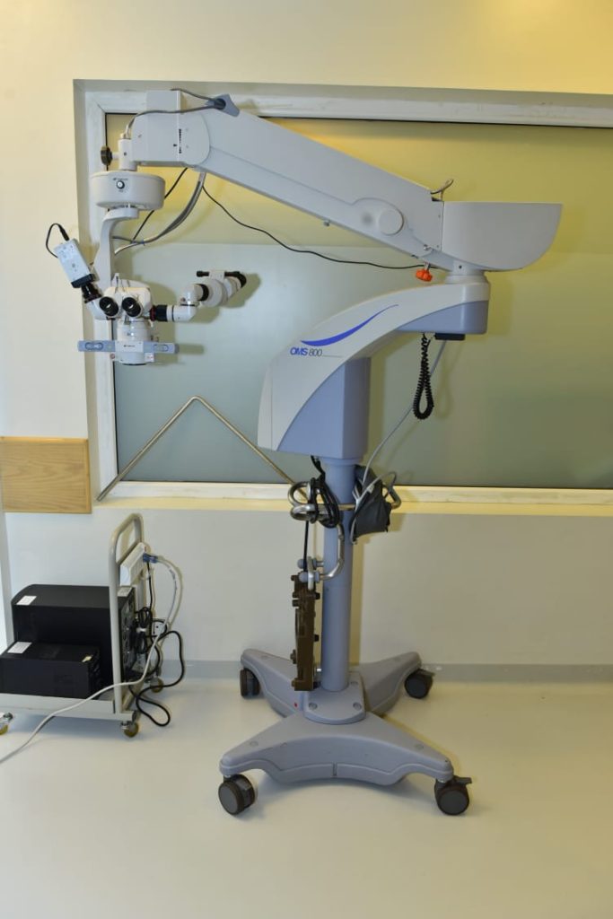
At The Eye Center, our 14 extremely well trained and highly experienced eye specialists and eye surgeons provide the complete range of laser and surgical eye procedures. We have a fully equipped operating suite that is dedicated to only eye surgeries and is maintained using the highest standards of sterilisation and according to the strictest international safety protocols.
We are fully equipped with a anterior ( cataract, cornea, refractive lens options, anterior segment surgery), posterior segment (retina and vitreous), trauma, strabismus and ocluloplastic surgery set up.
All patients and any attendants who may be accompanying the patient are adequately pre-screened for covid as to maintain a safe and covid free environment. All technical staff, nursing staff and all doctors are fully vaccinated and undergo periodic PCR testing.
Adherence to well thought out covid SOP’s ensure safety for patients and medical staff alike and assist in providing optimal surgical care to all our patients.
Corneal Hysteresis (CH) is an assessment of the cornea’s ability to absorb and dissipate energy and has been shown to be independently predictive of visual field progression in glaucoma.
Different from thickness or topography, which are geometrical attributes, Corneal Hysteresis is a tissue property.
A confident glaucoma risk assessment can be made with Corneal Hysteresis. Only Ocular Response Analyzer® G3 measuresCorneal Hysteresis and Corneal Compensated IOP (IOPcc) using patented technology to assess the unique corneal biomechanical properties of your patient.
Corneal Hysteresis has shown to be an independent risk factor and more predictive of glaucoma development and progression than CCT or IOP. Using biomechanics, IOPcc is less influenced by corneal properties than Goldmann applanation tonometry. Clinical research shows compelling evidence that CH is a powerful asset for assessing patients at high risk for developing glaucoma and disease progression.
CH is an important factor in determining the risk of disease progression not only in open angle glaucoma but also in normotensive glaucoma.
IOPcc given below is an important measure of true intraocular pressure in the post refractive surgery population as it is unaffected by changes in corneal thickness.
Biometry is the method of using mathematical formulas to determine the refractive power of the eye. The refractive power of the human eye depends on the cornea, the lens, the ocular media and the axial length of the eye.
It is extremely important to know the exact measurements of different structures as well as the inter structure distances as these measurements of the eye are essential when planning for cataract surgery.
This biometry and specially accurate biometry with using formulas that employ artificial intelligence allow eye surgeons to pick the absolute best possible lens for an individual patient that may need to be implanted after cataract surgery.
There are multiple calculations or equations that can be used to derive the power of an artificial or intraocular lens that should be used in a particular patient but all these the equations are dependent on getting accurate parameters that are entered into the equations.
This is possible using modern optical rather than ultrasound based biometers. At The Eye Center we use the Haag Streit Lenstar LS 900 which allows us to make calculations in conditions such as pseudophakia, in the paediatric age group, after posterior segment or retinal surgery as well as after refractive surgery such as LASIK.
Lenstar 900 is world recognised in the performance of highly accurate biometry.
Corneal pachymetry
Corneal pachymetry is the ability to measure the thickness of the human cornea. A pachymeter is a medical device used to measure the thickness of the cornea in the human eye.
This is an essential piece of information required for many different kinds of surgical procedures but particularly so for procedures such as LASIK or other refractive surgical procedures.
It is also used in the planning of cataract surgery as well as other anterior segment operations.
Pachymetry also allows us to determine an important risk factor in the development of glaucoma.
Digital Fundus Flourescein Angiography has contributed to our understanding of many different disease processes in the eye involving the retina and the choroid.
FFA as it is commonly know is the most preferred method to image both the retinal as well as the choroidal circulation. Fundus Flourescein Angiography is an essential tool used by many ophthalmologists to diagnose a variety of common and rare retinal diseases and determining and using the pattern of images obtained as proof of disease and in coming up with the best possible treatment protocols.
The test involves dilation of the pupil and after using a small test administering a larger dose of the actual dye in a vein of the arm. This dye is different from iodinated dye which then goes into the circulation of the human body and then goes into the circulation of the eye.
Multiple times pictures of the eye are then taken in quick succession using a specialized digital camera and filters.
The different patterns of circulation seen allow different disease processes to be picked up. The test is extremely important in diseases such as a diabetes diabetes macular degeneration as well as in many inborn or acquired retinal and choroid diseases.
Abnormal patterns in angiography are seen either as blocking effect You can also see leakage of the dye, staining of tissue structures in the retina, pooling or colllection of dye in discrete locations, and transmission defects.
The test is quite painless and is done in the clinic by extremely skilled technicians in the presence of a qualified medical doctor. Images acquired as a result of this test are critical in the accurate diagnosis and construction of treatment protocols for a patient as well as in their follow-up.
The test is painless and is done in the clinic with the patient in an upright seated position.
