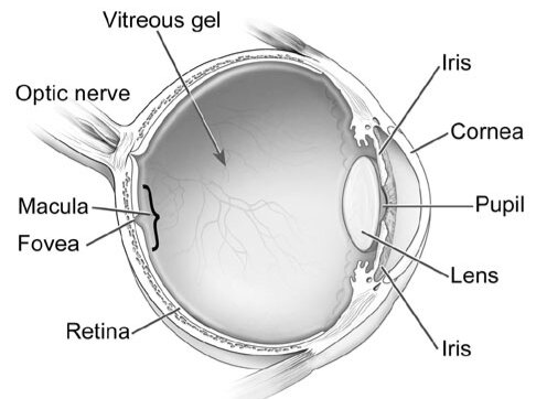
The vitreous, a material, fills the centre of the eye. This clear, gel-like substance is firmly attached to the retina and macula in the healthy, youthful eye by millions of tiny fibres. The vitreous shrinks and pulls away from the retina as the eye ages or as a result of an eye condition. Over time, the vitreous and retina totally separate which is known as a posterior vitreous detachment (PVD). It is typically a natural component of ageing. Most people experience it by the age of 70.
Some PVD sufferers experience partial vitreous detachment. The macula, which is the core of the retina, still has some vitreous attached to it. Vitreomacular Traction is brought on by the vitreous pulling and tugging on the macula (VMT). If addressed, this can harm the macula and impair vision.
A person’s chance of developing VMT may increase if they have certain eye conditions, such as high myopia (extreme nearsightedness), age-related macular degeneration (AMD), diabetic eye disease, retinal vein occlusion, or a disease that affects the blood vessels in the rear of the eye (a blockage of veins in the retina)
Additionally, taking pilocarpine to treat presbyopia, an age-related fuzzy near vision, can raise your risk of VMT.
The most typical signs of vitreomacular traction (VMT) include experiencing flashes of light in your vision, viewing objects as smaller than they actually are, and having distorted vision that causes a grid of straight lines to appear wavy, hazy, or blank.
These signs could potentially point to another eye condition. This is why it is crucial to get checked out by an ophthalmologist as soon as you start experiencing any of these symptoms.
Your ophthalmologist must examine the inside of your eye to make a diagnosis of VMT. One or more of these tests could be used to determine this.
The most frequent test ophthalmologists employ to detect VMT is optical coherence tomography (OCT), and fluorescein angiography is an imaging test that uses a specific camera to capture pictures of the retina. These tests aid in determining whether you have VMT. They can also inform your doctor of potential treatment needs.
There are typically three treatment options available after a vitreomacular traction (VMT) diagnosis. The observation or “wait and see” approach is when treatment might not be necessary if your VMT is minimal and not interfering with your vision. You and your ophthalmologist may elect to monitor (watch) the condition with follow-up visits since some cases of VMT will resolve on their own. Additionally, you will be required to use an Amsler grid at home every day to check your eyesight. Your ophthalmologist will go over how to use the Amsler grid correctly.
Surgery. When VMT is severe, it can cause retinal disorders that can impair vision, including cystoid macular edoema (swelling of the macula), macular pucker (when macular scar tissue builds up and distorts vision), and macular holes (when tugging of the vitreous creates a hole in the macula).
In these situations, a vitrectomy treatment may be suggested to return the macula to its normal (flat-lying) shape. The surgeon removes the vitreous from the eye with the help of microscopic devices and replaces it with saline solution. Under a microscope, any scar tissue on the macula is also removed using specialised tools. By doing this, the traction that is harming the macula is released.
Medication. Some individuals with severe VMT may not be suitable candidates for vitrectomy surgery. These people can receive treatment with a drug called ocriplasmin. The medication dissolves the microscopic protein fibres that link the vitreous with the macula and is injected intravitreally (injection into the centre of the eye). In order to relieve the traction, your ophthalmologist may also inject a gas bubble and instruct you to adjust your head in a particular way.
At The Eye Center- Dr. Mahnaz Naveed Shah & Associates our team of eight ophthalmology subspecialists/ eye specialists, eye surgeons who are considered amongst the very best eye specialists in Karachi and in Pakistan, have the diagnostic and treatment capabilities to treat from the simplest to the most complex patients. We work hard to provide our patients with the best possible medical and surgical eye care, in a state of the art purpose built eye care facility. We offer the entire array of medical, laser and surgical treatments to help provide patients the best possible care in the most efficient, safe and ethical manner.
If you need an appointment, please contact us at 03041119544 during our working hours or leave us a WhatsApp message at +923028291799 and someone will connect with you. Walk-in appointments are also available for emergencies. We can also be reached through our web portal on www.surgicaleyecenter.org

